
![]() A Simple Clinical Test For Severe Vitamin D Deficiencyitamin D is required for the correct metabolism of calcium. In this role, vitamin D coordinates with a number of other hormones, including parathyroid hormone and calcitonin, to regulate both calcium and phosphate levels in the blood. Generally calcitonin acts to lower plasma calcium levels and parathyroid hormone acts to raise plasma calcium levels. This system of hormonal interaction works effectively as long as adequate vitamin D is present. As calcium intake falls, the synthesis and release of parathyroid hormone increases. Parathyroid hormone acts to increase plasma calcium levels by increasing synthesis of 1,25-dihydroxyvitamin D, a metabolite of 25-hydroxyvitamin D. This then stimulates the reabsorption of calcium from the kidney and stimulates calcium uptake from the gut. However, in the vitamin D deficient state, the synthesis of 1,25-dihydroxyvitamin D cannot occur and so reabsorption and absorption of calcium from the kidney and gut, respectively, cannot be stimulated.
A Simple Clinical Test For Severe Vitamin D Deficiencyitamin D is required for the correct metabolism of calcium. In this role, vitamin D coordinates with a number of other hormones, including parathyroid hormone and calcitonin, to regulate both calcium and phosphate levels in the blood. Generally calcitonin acts to lower plasma calcium levels and parathyroid hormone acts to raise plasma calcium levels. This system of hormonal interaction works effectively as long as adequate vitamin D is present. As calcium intake falls, the synthesis and release of parathyroid hormone increases. Parathyroid hormone acts to increase plasma calcium levels by increasing synthesis of 1,25-dihydroxyvitamin D, a metabolite of 25-hydroxyvitamin D. This then stimulates the reabsorption of calcium from the kidney and stimulates calcium uptake from the gut. However, in the vitamin D deficient state, the synthesis of 1,25-dihydroxyvitamin D cannot occur and so reabsorption and absorption of calcium from the kidney and gut, respectively, cannot be stimulated.
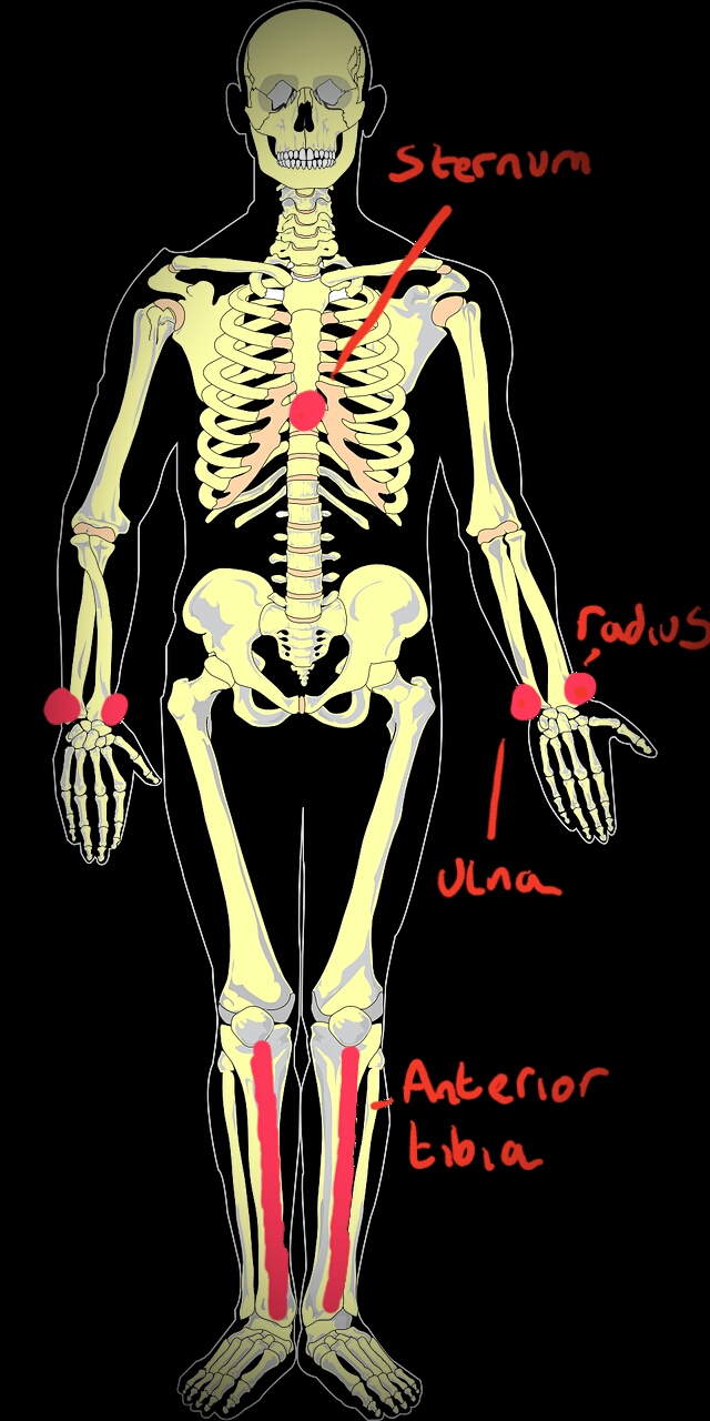
Minimum pressure with a thumb on the bottom part of the sternum, the styloid process of the ulna and radius in the lower arm, or the anterior tibia, is a clinical test for the presence of osteomalacia caused by a severe vitamin D deficiency. Vitamin D deficiency also causes muscle pain and weakness and muscle aches and pains in both children and adults. The combination of muscle weakness and weak bones leads to an increased risk of falls in the elderly and an increased risk that these will lead to serious damage to the bone. Clinically muscle and bone pain can be misdiagnosed as fibromyalgia.
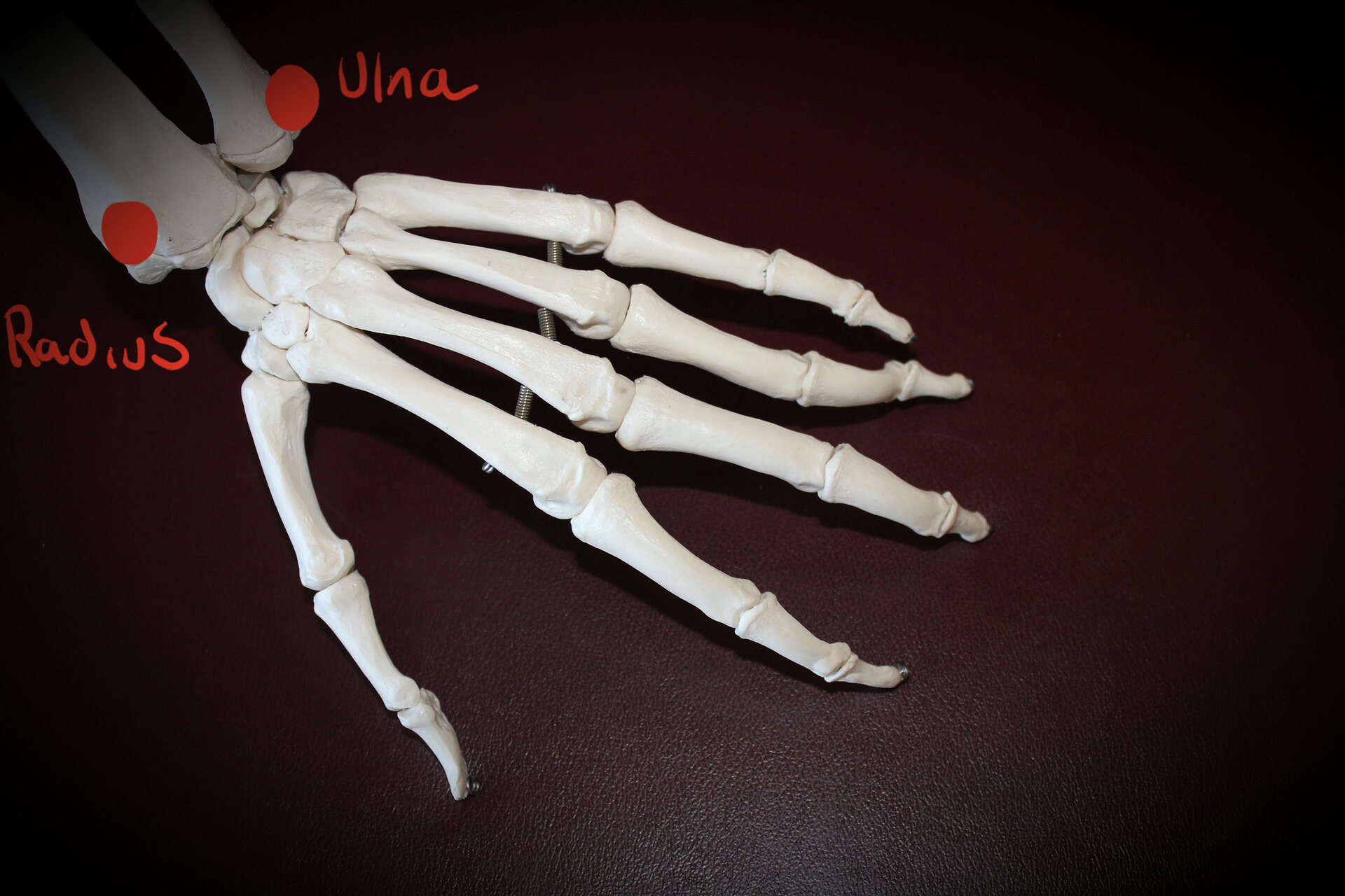
A simple clinical test for vitamin D deficiency can be application of a light pressure to the exposed parts of bones. The styloid process of the ulna and radius can be used for this purpose.
As a result of the lack of calcium absorption due to vitamin D deficiency, the skeleton starts to act as a surrogate donor of calcium to maintain blood levels through the activation of osteoclasts, and this leads to osteopenia and subsequently osteoporosis. Another effect of the raised levels of parathyroid hormone is the increased excretion of phosphate in the urine, and this can significantly reduce phosphate levels in the blood, reducing the availability of calcium phosphate to remineralise bone. However, the osteoblasts continue to deposit collagen matrix onto the periosteum of bone, but the low levels of calcium phosphate are not adequate to allow this matrix to mineralise. Instead it hydrates and expands, and this expansion causes an outward pressure that can lead to pain due to pressure on the periosteal covering. In this way vitamin D deficiency causes osteomalacia, which is characterised by bone strength deterioration and pain. Light pressure applied to exposed bone can be a clinical test for the presence of vitamin D deficiency.
Eat Well, Stay Healthy and Protect Yourself
RdB